
|
|||
|---|---|---|---|
|
|
Case report: nodular tuberculoid
leprosy Relato de caso: lepra tuberculoide nodular infantil en un paciente de 8 años de edad Relato de caso: hanseníase tuberculóide nodular da infância em uma paciente de 8 anos de idade |
|
|
|
Médico(as) Dermatologistas Hospital Eduardo de Menezes/FHEMIG, Belo Horizonte, MG (Brasil) |
Sandra Lyon | Adriana de Sousa Carneiro Alice Lage da Cunha | Marcela Fonseca Ladeira Renata de Sousa Carneiro | Elen Rose Teixeira Luciano José de Oliveira |
|
|
|
Abstract Nodular leprosy of childhood is a benign variation of tuberculoid leprosy, usually affecting children between the ages of 2 and 4 after long contact with virchowian patients. The case reported is that of a female eight-year-old patient, daughter of a virchowian father who presented nodular lesion, after one month of evolution. The histopatologic diagnosis confirmed the clinical hypothesis of nodular leprosy of childhood. Keywords: Leprosy. Nodular. Childhood.
Resumo Hanseníase nodular da infância é uma variante benigna da hanseníase tuberculóide, usualmente afetando crianças entre 2 e 4 anos de idade com contato prolongado com pacientes virchowianos. O caso relatado é de uma paciente do sexo feminino, 8 anos de idade, filha de um paciente com hanseníase virchowiana, que apresentava uma lesão nodular com um mês de evolução. O histopatológico confirmou a hipótese diagnóstica de hanseníase nodular da infância. Unitermos: Hanseníase tuberculóide nodular. Infância. Relato de caso.
Reception: 11/04/2015 - Acceptance: 02/07/2015
|
|||
|
|
EFDeportes.com, Revista Digital. Buenos Aires, Año 20, Nº 207, Agosto de 2015. http://www.efdeportes.com |
|
|
1 / 1
Introduction
Leprosy is an infectious disease caused by Mycobacterium leprae, a Gram-positive, acid-fast bacillus, with a tropism for the skin and peripheral nerves (Sampaio, 2007). When manifested in children under 15 years old, it reflects the intensity and long period of exposure to a high bacillary load. It represents, therefore, an important warning event, pointing to difficulties for controlling the disease (Sodré et al, 2007).
Nodular leprosy of childhood is a clinical variation of tuberculoid leprosy, which affects children exposed to a highly bacillipherous environment, as is the case with the children of virchowian patients (Fakhouri, Sotto, Manini & Margarido, 2003).
This report stresses the importance of an early clinical diagnosis for the nodular leprosy of childhood, thus interrupting the disease’s chain of transmission through epidemiologic research and eliminating leprosy as a public health issue.
Case report
Female patient, eight years old, with a pruriginous and not very painful lesion on the face, starting a month before. When that condition began, according to her relatives, the lesion presented pus, spontaneously evolving to a crusty aspect. No symptoms or related manifestations were reported. The child’s father had been under treatment for virchowian leprosy for a month (Figure 1).
Examination: papular, erythematous and crusty lesion, with a 3-mm diameter, at the left nasolabial fold (Figure 2). Sensitivity preserved with the 0.05gf monofilament. A scar from the BCG vaccine was also present.
A treatment with topical antibiotics was chosen, because of the secondary diagnostic hypothesis of pyoderma; however, no clinical improvements were observed, and the lesion enlarged. After a month of observation, a biopsy of the lesion under suspicion of nodular leprosy of childhood was performed and the treatment was initiated with children’s paucibacillary multidrug therapy. Mitsuda Test turned out positive (6 mm).
The histopathologic examination evidenced some epithelioid granulomas permeated by a moderate lymphocytic inflammatory infiltrate, irregularly distributed, which may correspond to nodular leprosy of childhood.
The patient proceeded with the treatment for leprosy without complications, evolving to the total disappearance of the lesion after the use of 6 monthly packs, in accordance with the treatment schedule for paucibacillary cases of leprosy (Figure 3).
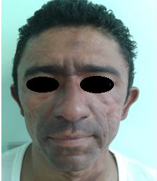
Figure 1. Child’s father under treatment for virchowian leprosy
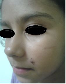
Figure 2. Child with papular, erythematous and crusty lesion
at the left nasolabial fold, before the beginning of the treatment
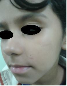
Figure 3. Child after treatment, total disappearance of the lesion
Discussion
Nodular leprosy of childhood is a clinically benign variation of tuberculoid leprosy, which affects children exposed to a highly bacillipherous environment, as is the case with the children of virchowian patients (Fakhouri et al, 2003). The lesions present themselves as small papules or nodules, which may be either brownish or reddish-brown, spontaneously healing and leaving a small area of atrophy (Sampaio, 2007). The lesions are usually few in number and are often isolated and located in areas of the skin in contact with the relatives’ infected areas (Souza Campos, 1937). There are no neural damages associated to this clinical form.
Despite its peculiar clinical evolution, the skin lesions’ histopathologic aspects are similar to those observed in the classic form of tuberculoid leprosy, showing tuberculoid granulomas which are well organized on the dermal layer (Fernandez, 1941). The smear microscopy is negative and the Mitsuda Test is positive (Sampaio, 2007).
Although nodular leprosy of childhood tends to heal spontaneously, some authors defend its early treatment. In Brazil, the Ministry of Health inserts nodular leprosy of childhood in the category of tuberculoid and advocates the treatment with 6 doses of paucibacillary multidrug therapy, consisting of 6 doses of dapsone and rifampicin for a period of up to 9 months (Ministério da Saúde, 2002).
Bibliography
-
Fakhouri, R., Sotto, M. N., Manini, M. I., & Margarido, L. C. (2003). Nodular leprosy of childhood and tuberculoid leprosy: a comparative, morphologic, immunopathologic and quantitative study of skin tissue reaction. International journal of leprosy and other mycobacterial diseases, 71(3), 218-226.
-
Fernandez, José M. M. (1941). Cicatriz residual da lepra tuberculóide infantil. Rev Bras Leprol; 9(4): 337-350.
-
Ministério da Saúde. Secretaria de Políticas de Saúde. Departamento de Atenção Básica. (2002). Guia para o Controle da Hanseníase. Brasília: Ministério da Saúde. Brasil.
-
Sampaio, Sebastião A.P. (2007). Dermatologia. 3ª Edição revista e ampliada. São Paulo: Artes Médicas.
-
Sodré, J.L., Nery, J.A.C., Carocha, A.P.G., Mendes, C.V., Rehfeldt, F.VS., Leitão, P.C. et al. (2007). Importância epidemiológica do diagnóstico clínico precoce da hanseníase nodular infantil. Hansen. Int;32(Special).
-
Souza Campos, N. (1937). Aspects cliniques de la lèpre tuberculoide chez l'enfant. Rev. Bras. Lepr. 5 (n.° especial), 33-113.
Another articles in English
|
|

Búsqueda personalizada
|
|---|---|
|
EFDeportes.com, Revista Digital · Año 20 · N° 207 | Buenos Aires,
Agosto de 2015 |
|